
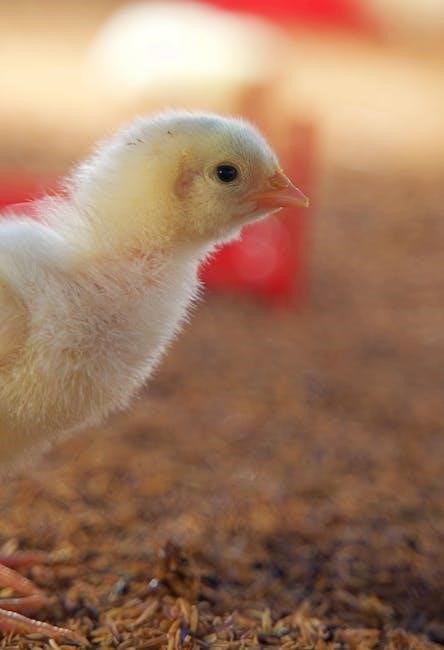
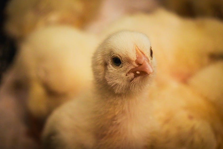
Chick embryo development, a fascinating biological process, unfolds over 21 days, offering a unique model for studying vertebrate development.
Detailed PDF guides and diagrams meticulously chart these stages, from initial cell division to hatching, providing invaluable insights for researchers and educators.

Overview of the 21-Day Incubation Period
The 21-day incubation period of the chick embryo is a remarkably organized sequence of events, meticulously documented in numerous chick embryo development stages PDF resources. Initially, the first few days witness rapid cell division and the formation of essential embryonic layers. Days 4-7 mark crucial organogenesis, including limb bud development and the beginnings of a beating heart.
As development progresses (Days 8-14), distinct features like feathers and the beak begin to form, alongside skeletal ossification. The final week (Days 15-21) focuses on yolk sac absorption, positioning for hatching, and the dramatic processes of internal and external pipping, culminating in the chick’s emergence. These PDF guides visually represent these stages, offering a comprehensive understanding of this complex developmental timeline, essential for both research and educational purposes.
Importance of Studying Chick Embryo Development
Studying chick embryo development is profoundly valuable due to its accessibility and similarities to other vertebrate species, including humans. Detailed chick embryo development stages PDF resources facilitate research into fundamental biological processes like organogenesis and cell differentiation. The transparent eggshell allows for non-invasive observation, making it an ideal ex-ovo model for studying angiogenesis and vasculogenesis, as highlighted in recent publications.
Furthermore, the relatively short incubation period and ease of manipulation make it a powerful tool for investigating developmental abnormalities and testing the effects of various interventions. Access to comprehensive PDF guides detailing each developmental stage is crucial for accurate analysis and interpretation of experimental results, advancing our understanding of vertebrate embryology and potential applications in regenerative medicine.

Early Stages of Development (Days 1-3)
Days 1-3 witness initial cell division, blastodisc formation, and the emergence of the area opaca and pellucida, as detailed in PDF guides.
Day 1: Initial Cell Division and Blastodisc Formation
Day 1 of chick embryo development marks the commencement of a remarkable journey, meticulously documented in various PDF resources detailing embryological stages. Initially, the single-celled zygote undergoes rapid cell division, a process known as cleavage. These divisions occur without significant growth in overall size, resulting in an increasing number of smaller cells.
This early cellular activity leads to the formation of a small, pale disc-shaped structure on the yolk surface called the blastodisc. This blastodisc, composed of approximately 5,000 cells, represents the precursor to the future embryo. Detailed diagrams within embryology PDF guides illustrate the arrangement of these cells and the initial polarization that begins to establish the body axes. The blastodisc isn’t yet fully organized, but it contains the potential for all future embryonic structures. Understanding this initial stage, as presented in comprehensive PDF references, is crucial for comprehending subsequent developmental events.
Day 2: Area Opaca and Area Pellucida Development
Day 2 witnesses a crucial differentiation within the blastodisc, clearly illustrated in chick embryo development PDF guides. The blastodisc now divides into two distinct regions: the area opaca and the area pellucida. The area opaca, centrally located, appears opaque due to its dense cellularity and will eventually contribute to the yolk sac. Surrounding it is the area pellucida, a clear, translucent region that harbors the cells destined to form the embryo proper.
These distinctions are visually detailed in embryological PDF resources, showcasing the developing organization. The area pellucida contains the embryonic and extraembryonic cells that will participate in gastrulation. Blood islands, precursors to the circulatory system, begin to form within the area pellucida. Studying these early morphological changes, as depicted in comprehensive PDF references, provides essential insight into the foundational steps of chick embryogenesis and the subsequent formation of vital organ systems.
Day 3: Primitive Streak Formation and Gastrulation Begins
Day 3 marks a pivotal moment in chick embryo development: the formation of the primitive streak, meticulously documented in chick embryo development PDF guides. This structure, appearing as a thickened line on the epiblast, initiates gastrulation – a fundamental process of cell rearrangement. Cells from the epiblast migrate through the primitive streak, establishing the three germ layers: ectoderm, mesoderm, and endoderm.
Detailed diagrams within embryological PDF resources illustrate this process, showing cells ingressing and spreading to form these layers. The primitive streak’s formation is driven by epithelial-to-mesenchymal transition (EMT), a key event highlighted in advanced PDF studies. This process is crucial for establishing the body plan and laying the foundation for organogenesis. Understanding the timing and mechanics of primitive streak formation, as presented in comprehensive PDF references, is essential for comprehending subsequent developmental events.

Heart Development – Initial Stages (HH Stages 3-10)
HH Stages 3-10, detailed in chick development PDF guides, showcase cardiac progenitor cell migration and primary heart field formation from the splanchnic mesoderm.
HH Stage 3: Cardiac Progenitor Cell Migration
HH Stage 3 marks a pivotal moment in cardiogenesis, comprehensively documented in chick embryo development PDF resources. At this stage, cardiac progenitor cells, originating within the epiblast, initiate a crucial process known as gastrulation. This involves an epithelial-to-mesenchymal transformation (EMT), a fundamental cellular shift enabling these cells to detach and begin their migration.
These migrating cells journey towards the anterior region of the embryo, ultimately forming the primary heart fields. These fields are derived from the splanchnic mesoderm, a key embryonic contributor to heart development. Detailed diagrams within these PDF guides visually illustrate this dynamic cellular movement and the subsequent organization of the progenitor cells. Understanding this initial migration is paramount to comprehending the subsequent formation and function of the developing heart, as outlined in research like Martinsen (2005).
HH Stage 4-10: Primary Heart Field Formation & Initial Heart Looping
Following the migration detailed in chick embryo development PDF guides, HH Stages 4 through 10 witness the consolidation of the primary heart field, originating from the splanchnic mesoderm. This field is a crucial early contributor to heart formation, as highlighted by Martinsen (2005). Simultaneously, a remarkable morphological event occurs: initial heart looping.
This looping, vividly illustrated in developmental diagrams found within these PDF resources, is a fundamental step in establishing the correct cardiac asymmetry and chamber positioning. The heart tube undergoes a characteristic bending process, setting the stage for future chamber development. These stages are also characterized by continued cellular differentiation and the beginnings of cardiac function. Understanding these processes, as detailed in comprehensive guides, is essential for studying congenital heart defects and normal cardiovascular development.

Mid-Stages of Development (Days 4-7)
PDF resources showcase rapid organogenesis during days 4-7, notably limb bud development and the enclosure of the heart within the thoracic cavity, resembling a bird.
Day 4-6: Organogenesis Begins – Limb Bud Development
Organogenesis, the formation of organs, commences dramatically between days 4 and 6 of chick embryo development. Detailed PDF guides visually document the emergence of distinct structures. Crucially, this period marks the initiation of limb bud development, representing the precursors to wings and legs. These buds, initially appearing as small swellings, rapidly elongate and differentiate.
The splanchnic mesoderm, a key embryonic contributor, actively participates in this process, alongside other tissues. Researchers utilize these developmental stages, as illustrated in various scientific diagrams available in PDF format, to study the intricate molecular mechanisms governing limb formation. Observing these changes ex-ovo allows for controlled experimentation and detailed analysis. The precision of these developmental events is remarkable, making the chick embryo a powerful model for understanding vertebrate embryology.
Day 7: Digit Formation and Heart Enclosure in Thoracic Cavity
By day 7, significant advancements are visible in the developing chick embryo, meticulously documented in comprehensive PDF guides. Most notably, digits begin to appear on both the developing wings and feet, marking a crucial step in limb morphogenesis. These early digits, though rudimentary, foreshadow the future functional limbs. Simultaneously, a vital internal event occurs: the heart becomes fully enclosed within the protective thoracic cavity.
This enclosure safeguards the rapidly developing heart, allowing for continued, robust function. Scientific diagrams, often found in PDF resources, illustrate this anatomical shift. Continuous monitoring, utilizing ex-ovo models, is essential to track these changes. The coordinated development of both external limbs and internal organs highlights the intricate orchestration of embryological processes, making the chick embryo an invaluable research subject.
Development of the Splanchnic Mesoderm
The splanchnic mesoderm, a crucial embryonic contributor, plays a pivotal role in heart development, as detailed in numerous PDF guides on chick embryology. This mesodermal layer gives rise to the primary, secondary heart fields, and the proepicardium – three major components essential for cardiac formation. Detailed diagrams within these resources illustrate the origin and migration of cells.
Cardiac progenitor cells, originating in the epiblast (around HH stage 3), undergo epithelial-to-mesenchymal transformation (EMT) and migrate to form this splanchnic mesoderm. This process, thoroughly documented in research papers and PDF references, is fundamental to establishing the foundations of the circulatory system. Furthermore, extracardiac contributors like the cardiac neural crest also significantly contribute to the developing heart, showcasing the complexity of this process.
Later Stages of Development (Days 8-14)
PDF guides reveal that days 8-14 witness significant advancements: feather and beak development begin, alongside skeletal ossification and continued organ refinement within the embryo.
Day 8-10: Feather and Beak Development
PDF resources detailing chick embryogenesis highlight days 8 through 10 as a period of dramatic morphological change. This is when the first visible signs of feather and feather tracts emerge, marking the beginnings of the chick’s future plumage. These structures aren’t fully formed yet, but their placement is clearly discernible. Simultaneously, the beak begins to harden, transitioning from a soft, cartilaginous structure to a more defined shape.
Detailed diagrams within these PDF guides illustrate the progressive development of both features. The feather follicles are initiated, and the beak undergoes keratinization, a process crucial for its strengthening. Observing these changes requires careful examination, often facilitated by ex-ovo models described in available literature. This stage is pivotal for understanding avian morphology and the genetic controls governing these developmental processes, as outlined in various research papers.
Day 11-14: Ossification of Skeleton and Further Organ Refinement
PDF guides dedicated to chick embryo development showcase days 11-14 as a critical period for skeletal maturation. Ossification, the process of bone formation, becomes increasingly prominent, with cartilage gradually replaced by hard bone tissue. This is visually documented in detailed diagrams found within these resources. Simultaneously, ongoing organogenesis leads to further refinement of internal structures, enhancing their functionality.
These PDFs often include illustrations demonstrating the progression of ossification in limbs, the skull, and the vertebral column. Organ systems, including the digestive and respiratory tracts, continue to develop and differentiate. Researchers utilize ex-ovo models, as described in scientific literature, to observe these intricate changes. Understanding this stage is crucial for studying developmental biology and potential congenital abnormalities, as highlighted in key research papers.
Advanced Development & Hatching (Days 15-21)
PDF resources detail yolk sac absorption and positioning for hatching during days 15-21, culminating in internal and external pipping before the chick emerges.
Day 15-18: Yolk Sac Absorption and Positioning for Hatching
During days 15-18 of chick embryo development, a crucial phase unfolds, extensively documented in various PDF guides detailing embryological stages. The embryo actively absorbs the remaining yolk sac contents, providing essential nutrients for final growth and development. This process is vital for fueling the energy demands of the rapidly maturing chick.
Simultaneously, the embryo begins to orient itself within the egg, assuming a position optimal for hatching – typically head tucked under the right wing. This positioning is critical for successful pipping and emergence. Detailed diagrams within these PDF resources illustrate the anatomical changes occurring during this period, showcasing the shrinking yolk sac and the embryo’s shifting posture.
Furthermore, the allantois, responsible for waste storage, continues to regress. The chick prepares its beak for the first break in the shell, a process meticulously outlined in developmental charts. Observing these stages ex-ovo, as described in research papers, provides valuable insights into the complex choreography of hatching preparation.
Day 19-21: Internal Pipping, External Pipping, and Hatching
The final 72 hours of chick embryo development, comprehensively illustrated in PDF guides, mark the dramatic transition to hatching. Internal pipping, where the chick breaks into the air cell, initiates the process around day 19. This is followed by external pipping, the first visible crack in the shell, typically occurring on day 20 or 21.

Detailed PDF resources showcase the chick’s use of its egg tooth, a specialized structure aiding in shell penetration. Hatching itself is a strenuous process, requiring significant energy expenditure. The chick rotates within the egg, gradually chipping away at the shell until it emerges.
Researchers utilizing ex-ovo models, as detailed in scientific diagrams, can closely monitor these final stages. These PDF guides emphasize the importance of proper humidity and temperature during this period for successful hatching. Observing the complete 21-day journey, from initial cell division to emergence, provides a profound understanding of avian development.

Monitoring Chick Embryo Development
PDF guides and diagrams facilitate careful observation of developmental stages. Continuous monitoring, including ex-ovo models, is crucial for understanding embryonic progress and identifying anomalies.
Methods for Observing Development (Ex-Ovo Models)
Ex-ovo culture techniques represent a pivotal advancement in chick embryo research, allowing for detailed observation and manipulation outside the protective eggshell. This method, often detailed in comprehensive PDF guides on chick embryo development stages, involves carefully removing the embryo from its shell after a specific incubation period – typically around day 3-7 – and maintaining it in a controlled, artificial environment.
Researchers utilize specialized incubators mimicking the original egg’s temperature and humidity, alongside nutrient-rich media to sustain viability. Ex-ovo models enable direct visualization of organogenesis, cardiovascular development, and skeletal formation. Furthermore, they facilitate experimental interventions, such as gene editing or drug administration, to assess their impact on embryonic development. Detailed scientific diagrams, frequently found in accompanying PDF resources, illustrate the precise setup and monitoring procedures. This approach is invaluable for angiogenesis and vasculogenesis studies, as highlighted in recent publications, offering a powerful tool for understanding fundamental developmental processes.
Importance of Continuous Monitoring
Continuous monitoring is paramount throughout chick embryo development, as subtle changes can indicate critical developmental milestones or potential anomalies. Detailed PDF guides outlining chick embryo stages emphasize the need for regular observation, often utilizing techniques like windowing the eggshell or employing ex-ovo models for direct visualization.
Consistent tracking allows researchers to correlate morphological changes with specific developmental stages, ensuring accurate data collection and interpretation. The need for this vigilance is underscored by the rapid pace of organogenesis, particularly during days 4-7, and skeletal ossification from day 11 onwards. Any deviation from expected timelines, as illustrated in developmental diagrams within PDF resources, warrants further investigation. Monitoring also aids in assessing the efficacy of experimental interventions and identifying potential teratogenic effects, ultimately contributing to a deeper understanding of vertebrate embryology.
Resources for Further Study
PDF guides, scientific diagrams, and key research papers on chick embryo development are readily available online, offering comprehensive insights into these fascinating stages.
Available PDF Guides and Scientific Diagrams
Numerous PDF guides comprehensively document the 21-day chick embryo development process, offering detailed stage-by-stage breakdowns. These resources, often available from university extension services like Mississippi State and Texas A&M AgriLife, visually illustrate the embryo’s transformation from initial cell division to hatching.
Scientific diagrams, frequently accompanying research publications – such as those found on Wiley Online Library concerning heart development – provide precise anatomical representations at various hours of incubation. These diagrams are invaluable for understanding complex processes like gastrulation, organogenesis, and skeletal ossification.
Furthermore, resources detailing ex-ovo models, like those used for angiogenesis studies, often include illustrative diagrams showcasing developmental progress. These materials facilitate a deeper understanding of the observable changes throughout the chick embryo’s life cycle, aiding both research and educational pursuits.
Key Research Papers on Chick Embryo Development
Significant research utilizing the chick embryo model has yielded crucial insights into vertebrate development. Papers like Martinsen’s (2005) in Developmental Dynamics, accessible via Wiley Online Library, detail the intricate stages of heart embryology, focusing on splanchnic mesoderm contributions and potential cardiovascular malformations.
Studies examining the primary and secondary heart fields, alongside proepicardium development, provide a foundational understanding of cardiogenesis. Research exploring epithelial-to-mesenchymal transformation (EMT) during gastrulation, as highlighted by Garcia-Martinez and Schoenwolf (1993) and Cohen-Gould & Mikawa (1996), is also pivotal.
Publications detailing ex-ovo models and angiogenesis studies offer valuable methodologies and visual data, often presented with supporting diagrams, furthering our comprehension of embryonic development stages and vascularization processes.